Ultrasound examinations
Ultrasound
Specialised Doctors YESY
Types of Examinations Provided
At the Ultrasound Department of Diktaion Medical Center we offer an extensive range of diagnostic ultrasound services, covering a wide range of medical needs. Indicatively, the basic tests include:
Κωδ. GHS: 76700 (for Upper Abdomen) and 76856-RAD (for Lower Abdomen)
Upper and Lower Abdominal Ultrasound (Pelvic Ultrasound) is a non-invasive and painless diagnostic test that uses ultrasound to image internal organs of the abdominal and pelvic region, in order to evaluate their structure and function.
In particular, it includes:
➤ Ano Koelia:
- Liver, gallbladder and gallbladder: to detect stones, inflammation, liver lesions or cholelithiasis.
- Pancreas: to assess pancreatic inflammation or masses.
- Kidneys: for cysts, stones, hydronephrosis or other kidney diseases.
- Spleen: for an increase in size or other abnormal conditions.
- Large vessels: such as the abdominal aorta for aneurysm.
➤ Kato Kilia (Pyelos):
- Bladder: to detect stones, polyps, tumours or residual urine.
- Reproductive organs:
- In women: uterus, endometrium and ovaries, for cysts, fibroids, polycystic ovaries, etc.
- In men: prostate and seminal cysts, for hyperplasia or inflammation.
This test is particularly useful for investigating symptoms such as:
- Pain or bloating in the abdomen
- Hematuria or changes in urination
- Gastrointestinal or gynaecological disorders
- Preventive control
Κωδ. GHS: 76770
Ultrasound examination of the retroperitoneal cavity (Kidneys, Aorta, Lymph nodes) is a diagnostic imaging examination performed using ultrasound and aims to evaluate vital organs and structures located at the back of the abdominal cavity. In particular, it includes the examination of:
- Kidney: to detect conditions such as cysts, stones, hydronephrosis, inflammation or tumours.
- Aortic: to detect abdominal aortic aneurysms or other vascular abnormalities.
- Retroperitoneal lymph nodes: to detect lymphadenopathy, which may be associated with infections, inflammatory conditions or neoplasms.
It is a painless, safe and non-invasive test that is often used as a preventive measure or as part of a diagnostic test in cases of abdominal pain, hypertension, haematuria or unexplained weight loss.
Κωδ. GHS: 76536
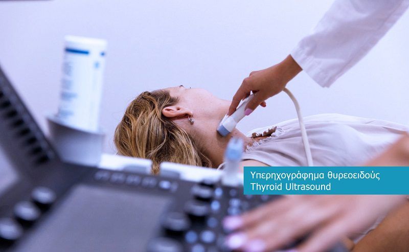
Ultrasound Head and Neck Soft Tissue (Thyroid, Parathyroid) is a painless, safe and non-invasive diagnostic test that uses high-resolution ultrasound for the imaging of anatomical structures in the neck area.
The purpose of the test is to evaluate:
➤ Thyroid gland:
- Detection of nodules or cysts
- Assessment of the morphology, size and perfusion of the gland
- Monitoring of known thyroid conditions such as thyroiditis, goitre or autoimmune diseases (e.g. Hashimoto’s)
- Supplementary testing in case of thyroid hormone disorders
➤ Parathyroid Adenoids:
- Detection of parathyroid adenomas or hyperplasia associated with hypercalcaemia and parathyroidism
➤ Other Soft Cervical Molecules:
- The test is recommended in people with:
- Palpable swelling in the cervix
- Abnormalities in thyroid or calcium blood tests
- Symptoms such as hoarseness, dysphagia or localised pain in the throat
- Family or personal history of endocrine disorders
Κωδ. GHS: CY242
Breast Ultrasound is a non-invasive, painless and safe diagnostic test that uses high frequency sound waves to visualize the soft tissues of the breasts.
It is an essential tool for the early diagnosis and monitoring of breast diseases, especially in young women or women with dense mammographic texture.
Breast ultrasound is used for:
- Investigation of palpable findings or symptoms such as pain, swelling or tenderness
- Distinction between solid and cystic lesions
- Identification of cysts, fibroadenomas or other benign morphomas
- Evaluation of suspicious lesions detected by mammography or clinical examination
- Biopsy guidance for taking a sample from suspicious areas
- Checking the axillary lymph nodes
The test is indicated in:
- Women under 40 years of age, as the first imaging method
- Women with dense breasts where mammography has limited sensitivity
- Women and men with palpable breast lesions or pain
- Screening, in combination or alternating with mammography, depending on age and individual history
Breast ultrasound can make a significant contribution to the early diagnosis of breast cancer and is often used as part of regular screening
Κωδ. GHS: 76870
Scrotal Ultrasound is a painless, safe and highly accurate diagnostic test that allows detailed imaging of the testicles, epididymis and other structures of the scrotal region.
The scrotal ultrasound is used for:
- Investigation of testicular pain or swelling
- Detection of testicular tumours or other masses
- Diagnosis of varicocele (enlargement of veins in the testicle), a common cause of infertility
- Detection of hydrocele (accumulation of fluid around the testicle)
- Diagnosis of testicular torsion, an urgent condition that requires immediate treatment
- Distinguishing between inflammation and other conditions, such as orheoepididymitis
- Evaluation of injuries in the scrotal region
- Infertility testing, combined with a spermogram
The examination is performed by placing the ultrasound sensor externally in the scrotum, without any discomfort for the patient, and is often accompanied by a Doppler test to assess blood flow.
It is recommended in cases of:
- Sudden or chronic pain in the testicles
- Swelling or mass palpator
- Injury
- Fertility Control
- Localised changes in the area
Κωδ. GHS: 76881
Ultrasound joint testing is a non-invasive, painless and dynamic diagnostic test used to visualize the soft tissue and functional status of one or more joints in the body.
The joint ultrasound can evaluate:
- Articular structures: such as synovial membrane, bursae, tendons and ligaments
- Existence of fluid or collection inside the joint (synovial effusion)
- Inflammatory lesions, e.g. in rheumatoid arthritis or gout
- Soft tissue injuries, such as tendonitis or tears
- Degenerative changes due to osteoarthritis
- Evaluation of inserts and periarticular cysts, such as Baker’s cysts
- Guiding injections or punctures under ultrasound guidance for precision
The test is applied to joints such as:
- Shoulder, for inspection of the rotor blade and subcrown pouch
- Knee, for fluid, meniscus tears or degeneration
- Hip, for hymenitis, collections, or injuries
- Podocnemic/carpal/metacarpal, for tendons, arthritis or injuries
- Small joints of hands/feet, in rheumatological patients
It is indicated in patients with:
- Pain, stiffness or swelling in a joint
- History of rheumatological or degenerative diseases
- Injury or suspected rupture of a tendon or ligament
- Need for paracentesis or local treatment
Joint ultrasound offers the possibility of real-time dynamic assessment, which makes it particularly useful for diagnosis and guiding treatment.
Κωδ. GHS: 93880
(TRIPLEX) Carotid and Vertebral Artery Ultrasound is a specialized, non-invasive and painless diagnostic test, which combines ultrasound imaging and Doppler blood flow monitoring.
It evaluates the function and morphology of the carotid and vertebral arteries, which carry blood to the brain.
The test is used to:
- Diagnosis of carotid artery stenoses or blockages (due to atheromatosis)
- Detection of atherosclerotic plaques, evaluation of their size and composition
- Estimating the risk of vascular stroke (VTE)
- Vertebrobasilar circulation control, especially in patients with dizziness, instability or fainting spells
- Monitoring patients with known vascular disease, hypertension, diabetes mellitus or hyperlipidemia
- Check-up after surgery or carotid angioplasty
Suitable for:
- People with a history of stroke or transient ischemic attack (TIA)
- People with cardiovascular risk factors: hypertension, diabetes, dyslipidemia, smoking
- Patients with dizziness, blurred vision, sudden loss of balance or fainting
- As a preventive measure, in elderly people or people with a family history of vascular disease
Carotid and vertebral artery TRIPLEX offers important information for diagnosis, prevention and treatment strategy in vascular and neurological problems.
Κωδ. GIS: 93925 (for Lower Extremities) and 93930 (for Upper Akron)
(TRIPLEX) Upper and Lower Limb Ultrasound is a modern, non-invasive and painless diagnostic test that combines ultrasound imaging with Doppler to assess blood flow in the veins and arteries of the upper or lower limbs.
The test is divided into:
➤ Venous Triplex:
- Checks for thrombosis (DVT), mainly in the lower limbs
- Evaluates venous insufficiency or varicose veins
- Records the functionality of the venous valves
- Used for preoperative testing or monitoring after surgery
➤ A rterial Triplex:
- Detects narrowing or blockages in the arteries of the limbs
- Assesses blood flow and pressure in cases of peripheral vasculopathy
- Helps diagnose intermittent claudication (pain when walking due to reduced blood flow)
- Monitor the progress of vascular surgery or angioplasty
Suitable for:
- People with swelling, pain, heaviness or tingling in the limbs
- Patients with varicose veins or a history of thrombosis
- Cases of suspected vascular occlusion or ischaemia
- People with diabetes mellitus, hypertension or arteriosclerosis
- Preventively, in patients with cardiovascular risk factors
TRIPLEX upper and lower limbs provides an accurate image of the circulation, contributing decisively to the diagnosis and treatment of vascular diseases.
Κωδ. GHS: 93970
The (TRIPLEX) Ultrasound Two-Way Scanning of the Limb Veins is a modern, non-invasive and completely safe diagnostic test, which combines:
- Gray-scale ultrasound (B-mode) for imaging of anatomical structures.
- Doppler scanning to evaluate the direction and velocity of venous flow.
- It evaluates in real time the function of the superficial and deep veins and the venous valves of the upper or lower limbs.
It is used for diagnosis:
- Deep vein thrombosis (DVT) – particularly in the lower limbs
- Superficial thrombophlebitis
- Chronic venous insufficiency and varicose veins
- Venous valve dysfunction (regurgitation/deficiency)
- Post-thrombotic syndrome
- Vascular complications after surgery or injury
- Preoperative vein mapping (e.g. in varicose vein or by-pass procedures)
It is indicated for people with:
- Pain, swelling, weight or burning sensation in the limbs
- Visible varicose veins or changes in skin color
- History of thrombosis or venous interventions
- Risk factors for thrombosis (prolonged immobility, surgery, malignancies, contraceptives)
- Symptoms indicative of venous stasis or insufficiency
Two-way venous scanning is the most reliable method for the diagnosis of venous diseases, as it allows accurate imaging and haemodynamic analysis of the venous circulation.
Κωδ. GIS: 93975
The (TRIPLEX) Scanning Ultrasound of the Scrotum – Abdomen is a specialized, non-invasive and painless diagnostic test, which is performed with dual-sided technology (B-mode + Doppler) and aims at the simultaneous evaluation of the arterial inflow and venous outflow of various anatomical areas, such as:
- The scrotum and its contents (testicles, epididymis, seminal vesicle, seminal vesicle)
- The abdomen, pelvic and lumbar region
- The retroperitoneal organs (such as kidneys, adrenal glands, lymph nodes, vessels)
The test is used to:
➤ Abdominal circulation:
- Diagnosis of varicocele (especially subclinical form)
- Evaluation of bidirectional venous outflow and reflux
- Testicular arterial adequacy test
- Infertility investigation in men
➤ Retroperitoneal and pelvic space:
- Detection of lymph node swellings, masses or vascular diseases
- Assessment of blood flow in major veins and arteries, such as the inferior vena cava, aorta, iliac arteries/veins
- Nutcracker syndrome or left renal vein compression syndrome screening
- Detection of venous insufficiencies or blockages affecting the circulation of the area
Indicated in cases:
- Male infertility or abnormal spermogram
- Clinical or subclinical varicocele
- Swelling or pain in the scrotum
- Symptoms of venous congestion in the pelvis
- Suspicious findings on previous ultrasounds or tests
- Pre-operative or post-operative control
- Unexplained haematuria or lumbar pain
This TRIPLEX provides a holistic view of the blood circulation in the scrotum and associated anatomical structures, and is particularly useful in cases of complex etiology or for detailed angiological mapping.

Why choose Diktaion Medical Center for your ultrasound examinations?
Cutting-edge technology
We use state-of-the-art ultrasound equipment, providing high-resolution images for accurate diagnosis.
Specialised Staff
The tests are performed by certified and experienced doctors and technologists, ensuring reliable results.
Safety and Quality
We adhere to strict health and safety protocols, while the continuous training of our staff guarantees a high level of service.
For more information or to schedule an appointment, please contact us. At Diktaion Medical Center, we are here to provide you with the best possible health care services.